
Orthopantomography | Radiology Reference Article
- Jun 18, 2025
- 4 min read
Orthopantomography, commonly known as OP or panoramic radiography, is a vital imaging technique in dentistry and maxillofacial radiology. It provides a wide and detailed view of the entire mouth, including teeth, jaws, and surrounding structures, all captured in one image. With advancements in radiology, understanding the significance and application of orthopantomography is increasingly important for healthcare professionals and patients alike.
What is Orthopantomography?
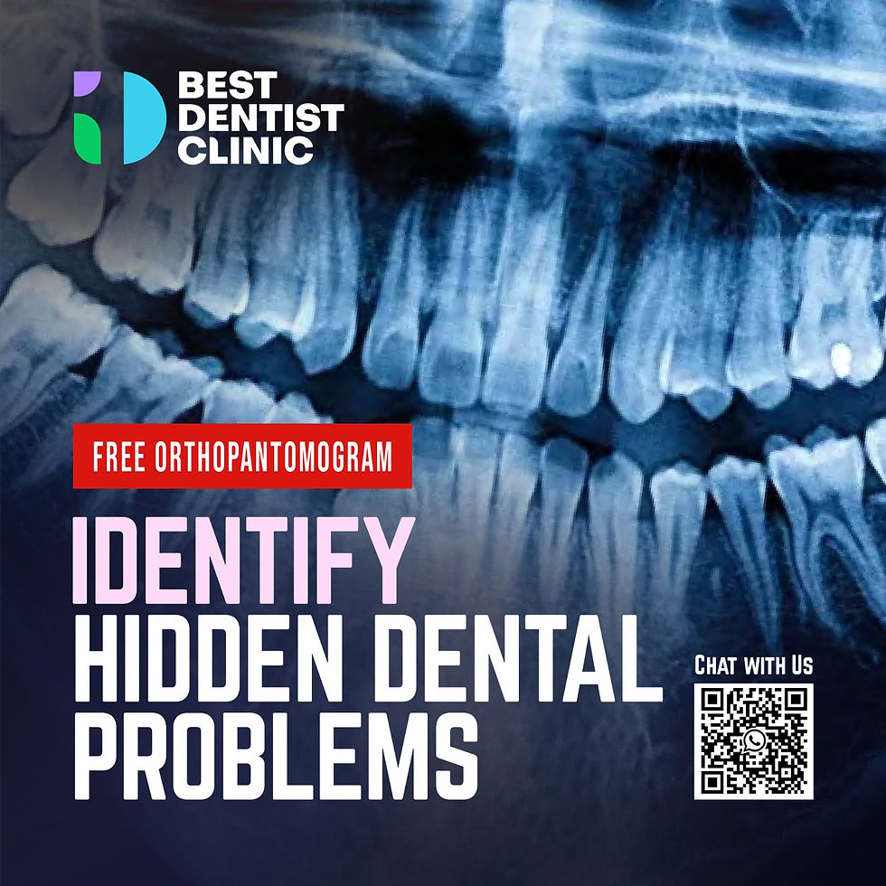
Orthopantomography is a specialized X-ray technique that captures a panoramic image of the dental arches and supporting structures. The equipment rotates around the patient’s head, emitting a beam of radiation that records data onto film or a digital sensor, producing a two-dimensional view of the oral cavity.
One key advantage of this technique is its efficiency. Traditional radiography often requires multiple images to obtain a complete picture, while OP allows for a single exposure that encompasses complete dental and skeletal anatomy. For example, an orthopantomogram can provide views of 32 teeth, both the upper and lower jaws, and their roots without the need for multiple X-rays, which not only saves time but also reduces patient exposure to radiation.
How Does Orthopantomography Work?
The process of orthopantomography involves positioning the patient in a standing or sitting posture. The patient must keep their head still as the X-ray machine rotates around them. This positioning is crucial for obtaining a clear and accurate image.
As the machine moves, it emits controlled radiation that captures the dental structures and surrounding tissues. This panoramic X-ray allows healthcare providers to identify various conditions such as tooth decay, jaw disorders, and the location of dental implants. For instance, studies show that OP can reveal the positioning of impacted wisdom teeth in 75% of cases, aiding in timely intervention.
Applications of Orthopantomography
Orthopantomography serves numerous purposes, particularly in dentistry and oral surgery. Some key applications include:
1. Dental Assessment
Orthopantomograms (OPGs) play a crucial role in dental evaluations. Dentists use OPGs to assess tooth alignment, detect impacted teeth, and plan for extractions or orthodontic treatments effectively. For instance, an OPG can reveal the presence of over-retained primary teeth, enabling prompt treatment.
2. Surgical Planning
When it comes to surgical procedures like wisdom teeth extraction, orthopantomography is instrumental. This imaging provides essential information on the spatial arrangement of teeth, roots, and adjacent nerves. A proper view increases the chances of a successful surgery, with studies indicating a reduction in surgical complications by up to 30% when using OP for planning.
3. Monitoring Diseases
In periodontology, OP images are essential for monitoring the progression of diseases such as periodontal disease. By providing an overview of bone levels and dental alignment, it allows dental professionals to make informed decisions. Research has shown that regular monitoring using OPGs can significantly improve treatment outcomes in patients with gum disease, with 85% of patients showing improvement after consistent examinations.
4. Implantology
In dental implant procedures, OP is a valuable tool. It helps assess bone quantity and quality at the implant site, which is vital for the successful integration of the implant. For example, a panoramic X-ray can identify areas with sufficient bone density, thereby increasing implant success rates by up to 95%.
5. TMJ Disorders
Orthopantomography aids in diagnosing temporomandibular joint (TMJ) disorders and provides insight into the anatomy of the joint and its relationship to the jaw and surrounding structures. This imaging can help identify issues affecting 10 million Americans, guiding appropriate treatment plans.
Benefits of Orthopantomography
The benefits of orthopantomography go beyond its diagnostic capability:
1. Reduced Radiation Exposure
A significant advantage of panoramic radiography is its relatively low radiation exposure compared to traditional X-ray methods. Research suggests that the radiation dose during an OP is about 10-20% lower than that of standard full mouth dental X-rays, making it a safer option for patients.
2. Patient Comfort
The non-invasive nature of OP typically results in less discomfort for patients during the procedure. The quick operation minimizes the time spent in the X-ray chair, helping to ease anxiety for those who may dread dental visits.
3. Comprehensive View
OP provides a broader view than traditional methods, allowing for a more thorough evaluation of both hard and soft tissues. This holistic imaging often leads to more precise diagnoses. In fact, studies have shown that dental professionals can make more informed decisions regarding treatment plans, with accuracy rates increasing by up to 25%.
Limitations of Orthopantomography
While orthopantomography is a powerful diagnostic tool, it does come with some limitations:
1. Image Distortion
The panoramic imaging process can sometimes result in distortion, causing magnification or misrepresentation of tooth positioning. This limitation may lead dentists to rely on additional imaging methods in complex cases.
2. Limited Detail
Compared to more advanced imaging techniques like cone-beam computed tomography (CBCT), OPGs may not provide the intricate detail needed for specific diagnoses, particularly when dealing with complex anatomical structures associated with 10% of dental cases.
3. Specificity of Indications
Orthopantomography is not suitable for every dental condition. Certain symptoms or issues may require alternative imaging methods to ensure accurate diagnosis and treatment planning.
The Future of Orthopantomography
Orthopantomography marks a significant advancement in dental radiography, offering efficiency alongside comprehensive diagnostic capabilities. By enabling a panoramic view of dental and jaw structures, OP has become an essential tool in modern dental practice.
Understanding both the strengths and limitations of this imaging technique is essential. Its applications in orthodontics, oral surgery, and general dentistry highlight its importance in providing effective patient care. As technology continues to evolve, orthopantomography will likely improve further, enhancing diagnostic capabilities and health outcomes for patients.
Effective interpretations paired with sound clinical judgment will help ensure the best results for those seeking comprehensive dental care. Recognizing the nuances of orthopantomography is crucial for healthcare practitioners committed to delivering precise and efficient dental solutions.


Comments